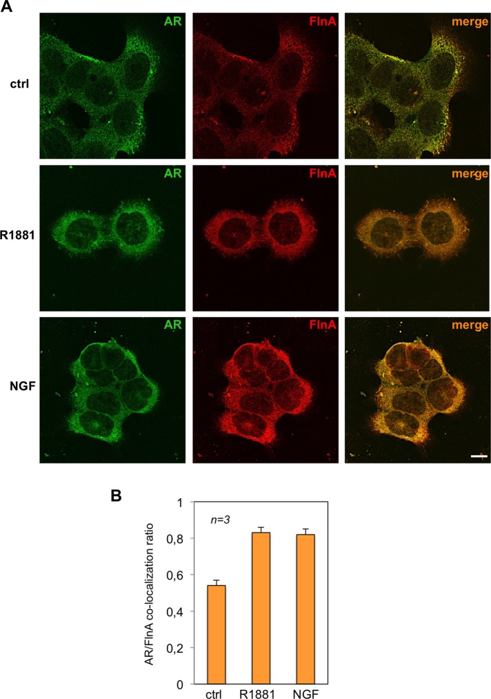
FIGURE 6:
R1881 or NGF induces extranuclear AR/FlnA complex in PC12 cells. Quiescent PC12 cells were left untreated (ctrl) or treated for 5 min with 10 nM R1881 (R1881) or 100 ng/ml NGF (NGF). (A) Cells on coverslips were visualized by IF for AR and Fln A. Images captured by confocal microscope show the staining of AR (green) and Fln A (red). Right, merged images. Confocal microscopy analysis is representative of three independent experiments. Bar, 10 μm. (B) Quantification and statistical analysis of these experiments. AR/FlnA colocalization ratio was calculated as described in Materials and Methods. Data from several independent experiments were analyzed. Means and SEM are shown; n represents the number of experiments. The statistical significance of results was also evaluated by paired t test. The difference in AR/FlnA colocalization ratio between unstimulated (ctrl) and R1881-stimulated cells was significant (p < 0.001). Also significant (p < 0.001) was the difference in AR/FlnA colocalization ratio between control (ctrl) and NGF-stimulated cells.

