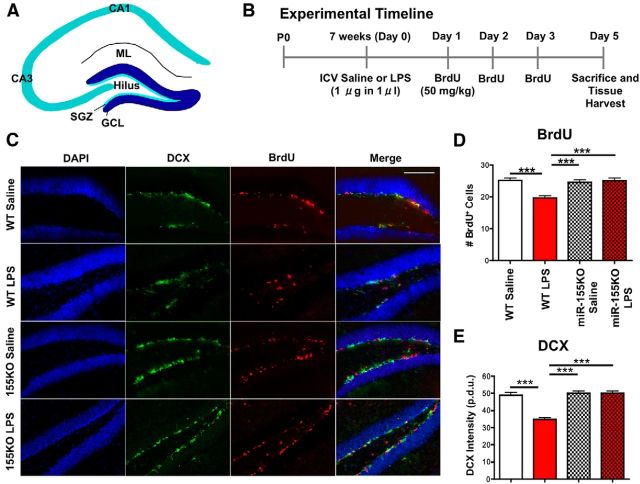
Figure 2.
Disruption of miR-155 leads to reversal of inflammation-induced decrease in NSC proliferation and neural differentiation in the DG. A, Diagram of the hippocampal DG. ML, Molecular layer; CA, cornu ammonis. B, Timeline of experimental design. C, Representative images of inflammation-induced changes in DCX (green; immature neurons), BrdU (red; proliferating cells), and DAPI (blue; nuclei) expression in the DG of WT and miR-155−/− (155KO) mice treated with saline or LPS (1 μg) intracerebroventricularly at 7 weeks of age. Scale bar, 100 μm. D, Quantification of total BrdU+ cells in the DG. E, DCX fluorescence intensity in the DG (procedure-defined units, p.d.u.). n = at least 10 images counted from 7 animals/ group. ***p < 0.001 by 1-way ANOVA and Tukey post hoc. Data are shown as mean ± SEM.

