Figure 5.
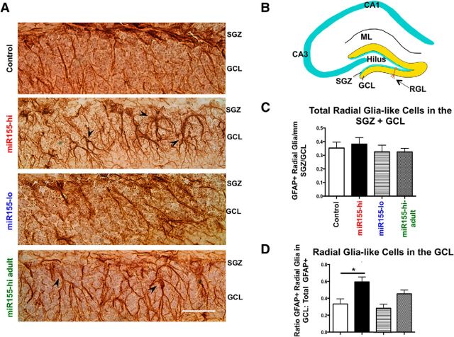
Elevated miR-155 leads to ectopic localization of radial-glia-like cells in the DG. A, Representative images of GFAP+ radial-glia-like cells in the SGZ and GCL in control, miR155-hi, miR155-lo, and miR155-hi-adult DG, showing their miR155-induced mislocalization. Original magnification = 40× objective. Scale bar, 100 μm. Arrowheads indicate ectopic GFAP+ radial-glia-like cell bodies. B, Diagram of DG showing radial-glia-like (type 1 progenitor) cells with normal cell body location in the SGZ. C, Total GFAP+ radial-glia-like cells in the SGZ per millimeter revealed no significant differences between groups. D, Ratio of GFAP+ cells in the SGZ versus GCL. n = 10–20 brain sections per group. Scale bar, 100 μm, Original objective = 40×. *p < 0.05 by 1-way ANOVA and Tukey post hoc. Data are shown as mean ± SEM.

