
Figure 13.
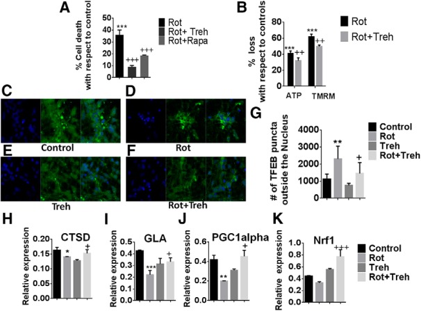
TFEB activation prevents losses in mitochondrial function and cell viability in mitochondrially compromised human iPSC-derived DAergic neurons. A, Measurement of cellular viability via MTT assay in iPSCs differentiated into DAergic cells treated with rotenone ± trehalose (Treh) or rapamycin (Rapa), expressed as % cell death versus untreated control. ***p < 0.001 versus untreated control. +++p < 0.001 versus rotenone alone. n = 3 per condition. B, Mitochondrial activity as monitored by ATP levels and TMRM fluorescence for measurement of mitochondrial membrane potential, expressed as % loss versus untreated control. ***p < 0.001 versus untreated control. ++p < 0.01 versus rotenone alone. n = 3 per condition. Activation of TFEB via measurement of cytoplasmic ICC in (C) untreated controls, (D) iPSC-derived neurons treated with rotenone (Rot) alone, (E) treatment with Treh alone, and (F) combined Rot/Treh. G, Quantitation reported as % TFEB puncta outside the nucleus. **p < 0.01 versus untreated control. +p < 0.05 versus Rot alone. TFEB target gene expression via RT-PCR for (H) cathepsin D (CTSD), (I) galactosidase α (GLA), (J) PGC1α, and (K) Nrf1. *p < 0.05 versus untreated control. **p < 0.01 versus untreated control. ***p < 0.001 versus untreated control. +p < 0.05 for Rot alone. +++p < 0.0001 for Rot alone. n = 3.