Figure 3.
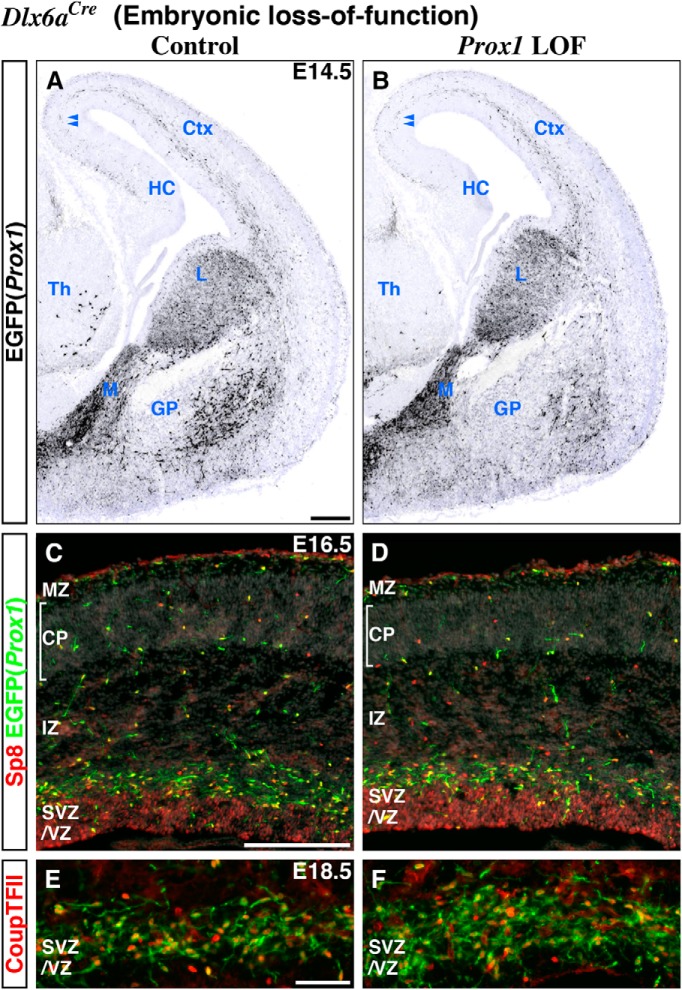
Embryonic (Dlx6a-Cre) Prox1 loss-of-function does not affect the interneuron precursors for their tangential migration from the CGE into the developing cortex as well as the expression of Sp8 and CoupTFII transcription factors. We performed embryonic (Dlx6a-Cre) control (A, C, E) and Prox1 loss-of-function (Prox1-C:EGFP/+ and /F) experiments (B, D, F). A, B, At E14.5, about the time the earliest cohort of CGE-derived interneuron precursors is first observed tangentially migrating through the intermediate zone (IZ) of the cortex (Miyoshi et al., 2010), we found no obvious differences between control and Prox1-null cells labeled with EGFP (shown in black) with regard to their migration pattern or distance from the CGE. Nuclear counter staining by DAPI is shown in blue. Double arrowheads indicate the CGE-derived interneuron precursors that have reached the border area between the hippocampus (HC) and cortex (Ctx). While control EGFP(Prox1)-labeled cells are positioned laterally to the developing globus pallidus (GP) and do not express CoupTFII (Nr2f2), in the Prox1 loss-of-function ventral telencephalon, very few EGFP(Prox1)-labeled cells are observed in the comparable domain. C, D, In addition to its expression within cortical progenitors, at E16.5, the Sp8 expression pattern within EGFP(Prox1)-labeled cells is comparable between the control and Prox1 loss-of-function cortex. E, F, Higher magnification pictures of the cortical subventricular/ventricular zones (SVZ/VZ) in Figure 2D and E. While there are significantly more EGFP-labeled cells found in the SVZ/VZ of the Prox1 loss-of-function cortex at E18.5 (see also Fig. 2F), the proportion of CoupTFII expression within EGFP(Prox1)-labeled cells is not changed. M, MGE; L, LGE; Th, thalamus; MZ, marginal zone; CP, cortical plate. Scale bars: A–D, 200 μm; E, F, 50 μm.

