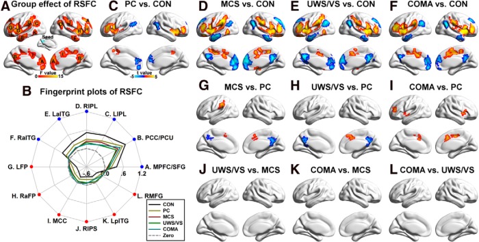
Figure 6.
Group differences in the PCC/PCU resting-state functional connectivity (RSFC). A, Main group effect of the PCC/PCU RSFC among the five groups (p < 0.05 corrected). B, Fingerprint plots of the mean fitted RSFCs of each region that showed a significant main effect across the five groups. Regions overlapped with the DMN are labeled in blue, while those overlapped with the SN/ECN are labeled in red. C–L, Post hoc comparisons of 10 pairs of groups (p < 0.05 corrected); two-sample t-maps between PC patients and CONs (C), MCS patients and CONs (D), UWS/VS patients and CONs (E), coma patients and CONs (F), MCS and PC patients (G), UWS/VS and PC patients (H), coma and PC patients (I), UWS/VS and MCS patients (J), coma and MCS patients (K), and between coma and UWS/VS patients (L) were shown. Of note, there was no significant difference between UWS/VS and MCS patients (J), between coma and MCS patients (K), or between coma and UWS/VS patients (L). MPFC/SFG, Medial prefrontal cortex extending to the bilateral superior frontal gyrus; LIPL, left inferior parietal lobule; RIPL, right inferior parietal lobule; LaITG, left anterior inferior temporal gyrus; RaITG, right anterior inferior temporal gyrus; LFP, left frontoparietal regions, including the intraparietal sulcus, insula, middle and inferior frontal gyrus; RaFP, right anterior regions of FP, including the insula, middle and inferior frontal gyrus; MCC, middle cingulate cortex; RIPS, right intraparietal sulcus; LpITG, left posterior temporal gyrus; RMFG, right middle frontal gyrus.

