Figure 1.

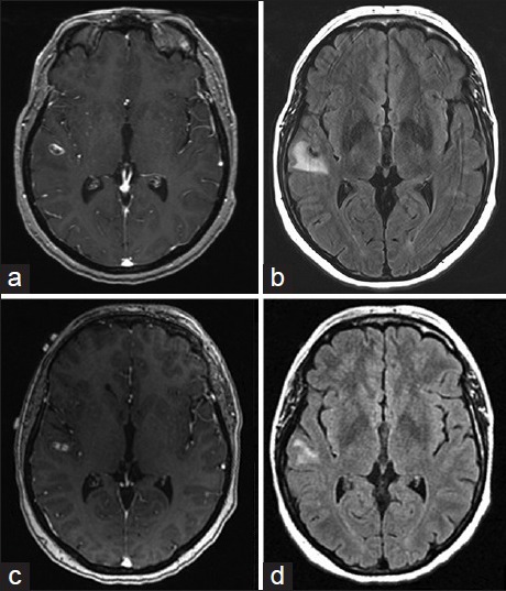
Magnetic resonance imaging brain with contrast and T2 fluid-attenuated inversion recovery (a) and (b) initial magnetic resonance imaging, (c) and (d) 1-month follow-up magnetic resonance imaging

Magnetic resonance imaging brain with contrast and T2 fluid-attenuated inversion recovery (a) and (b) initial magnetic resonance imaging, (c) and (d) 1-month follow-up magnetic resonance imaging