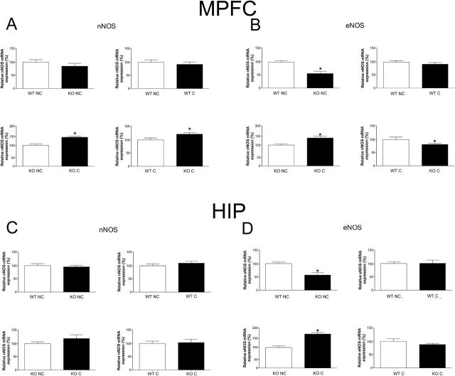
Figure 6.
Expression of neuronal nitric oxide synthase (nNOS) and endothelial nitric oxide synthase (eNOS) mRNA in the medial prefrontal cortex (MPFC) (A-B) and hippocampus (HIP) (C-D) of wild-type (WT) and inducible nitric oxide synthase (iNOS) KO mice. A) In the MPFC, conditioning (C) increase nNOS mRNA in KO mice compared with KO nonconditioned mice (NC), and KO C presented higher mRNA nNOS levels than WT C (n=7–8/group). B) In the MPFC, KO NC presented lower eNOS mRNA than WT NC, and conditioning increased eNOS mRNA in KO mice compared with KO NC, although KO C presented lower eNOS mRNA than WT C (n=6–8/group). C) In the HIP, there was no difference in the expression of nNOS mRNA. D) In the HIP, KO NC presented lower eNOS mRNA than WT NC, whereas conditioning increased eNOS mRNA in KO mice compared with KO NC (n=5–8/group). Results are expressed as percentage means±SEM of control values. Student’s t test, *P<.05.

