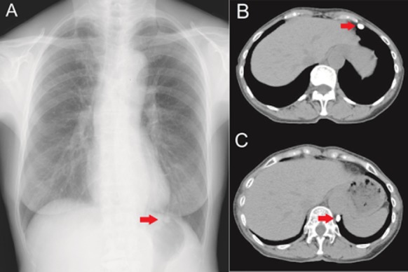
Figure 1.

(A) Chest radiograph shows a well-defined oval nodule (arrow) in the left lower lung field. (B) Chest computed tomography (CT) at the initial visit shows a well-defined, oval, calcified mass (arrow) on the ventral side of the left thoracic cavity. (C) Follow-up CT shows migration of the calcified mass (arrow) on the dorsal side of the left thoracic cavity.
