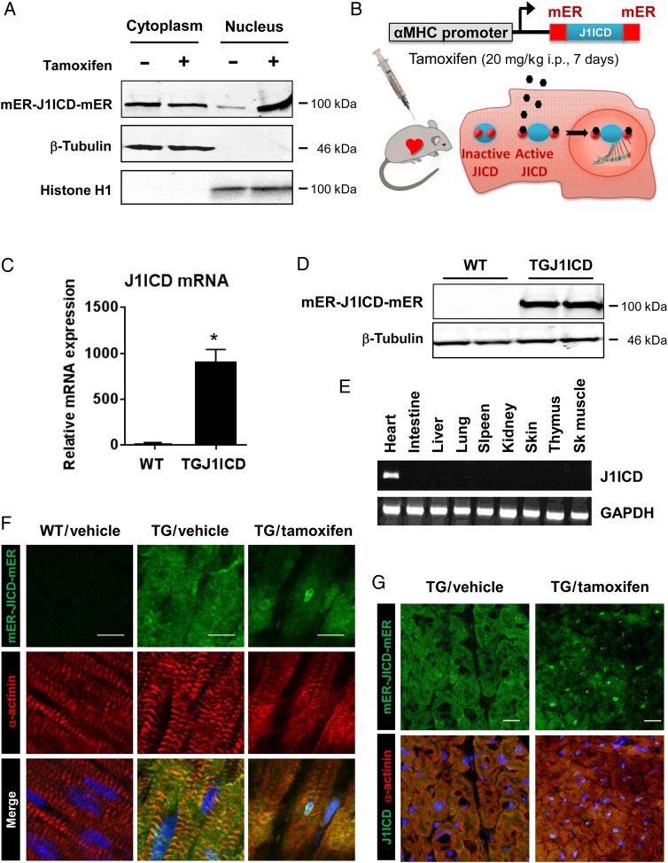
Figure 2.
TG mice with cardiac-specific expression and inducible activation of J1ICD. (A) HEK 293 cells transfected with pEF1α-mER-J1ICD-mER were treated with 4-OH-tamoxifen (1 µM) or DMSO. Cytoplasmic and nuclear fractions were separated and assessed for J1ICD expression by western blot. Anti-α-tubulin and anti-histone H1 immunoblots were performed as loading controls. (B) Schematic representation of the α-MHC:mER-J1ICD-mER cDNA used to generate TGJ1ICD mice (top). In the absence of tamoxifen, the mER-J1ICD-mER fusion protein forms a complex with HSP proteins and is sequestered in the cytosol. Tamoxifen injections release mER-J1ICD-mER and induce its nuclear translocation (bottom). (C) Quantitative RT-PCR analysis of the transgene expression. Results were normalized to endogenous Gapdh and Jagged1 expression and were expressed as mean ± SEM (n = 10–15 mice per group; *P ≤ 0.01). (D) Anti-Jagged1 immunoblot performed on whole heart extracts from WT or TGJ1ICD mice. (E) RT-PCR specific for the transgene and Gapdh gene were performed on different organs of TGJ1ICD mice to show the cardiac-specific expression of the transgene. (F and G) Heart sections from WT or TGJ1ICD mice, treated with vehicle or tamoxifen for 48 h, were immunostained with anti-Esr1 antibody, which recognizes the mER motif of the transgene (green) and anti-α-sarcomeric-actinin antibody (red). Nuclei were stained with DAPI (blue). Scale bar, 10 µm (F) and 20 µm (G). Images in F and G are representative of at least five mice per group.

