Figure 3.
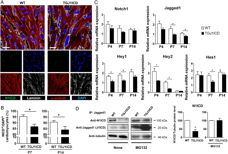
Notch activation is decreased in neonatal TGJ1ICD mice. WT and TGJ1ICD neonatal mice treated daily with tamoxifen were sacrificed at postnatal days P0, P4, P7, and P14. (A) Confocal images of heart sections immunostained against N1ICD (green), laminin (red), and α-sarcomeric-actinin (white). Nuclei were stained with DAPI (blue). Scale bar, 20 µm. (B) The number of NICD-positive cardiomyocytes at P7 and P14 was quantified on images depicted in A, and expressed as % of total DAPI-positive cardiomyocytes (n = 4–6 mice per group). (C) RNA expression of Notch target genes was analysed by quantitative RT-PCR. Results, normalized to Gapdh, represent fold change relative to the WT group at P0 and are expressed as mean ± SEM (n = 4 mice per group and per time point; *P ≤ 0.05). (D) Tamoxifen-treated mice were injected or not with MG132 from P9 to P14. J1ICD immunoprecipitation was performed using anti-Jagged1 antibody on whole heart extracts. Immunoprecipitates were immunoblotted with anti-N1ICD, anti-Jagged1 to detect J1ICD, and anti-tubulin antibodies. A quantification of N1ICD band intensity relative to tubulin is shown on the right panel (n = 5–6 mice per group). Bar graphs are mean ± SEM. *P ≤ 0.05 vs. WT.

