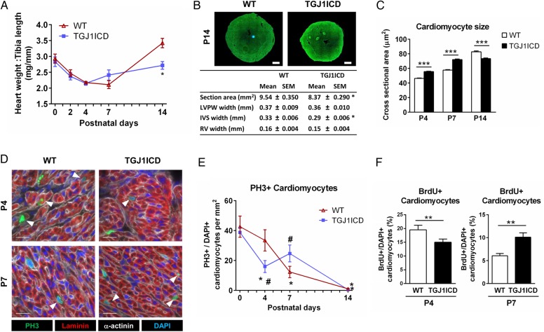
Figure 4.
Altered cardiomyocyte maturation in neonatal TGJ1ICD mice following J1ICD activation. TGJ1ICD neonatal mice treated daily with tamoxifen were sacrificed at postnatal days P0, P2, P4, P7, and P14. (A) The ventricular weight to tibia length ratio of WT and TGJ1ICD mice. (B) Heart sections of 2-week-old mice (top) were used to measure cardiac dimensions (bottom). LVPW: left ventricular posterior wall; IVS: interventricular septum; RV: right ventricle. Scale bar, 0.5 mm. (C) Cross-sectional areas of ≥1000 myocytes per group were measured on laminin-stained heart sections. B and C; n = 6 mice per group. *P ≤ 0.05, ***P ≤ 0.01 vs. WT. (D) Heart sections were immunostained against PH3 (green), α-sarcomeric-actinin (red), and laminin (white). Nuclei were stained with DAPI (blue). Arrows indicate PH3-positive cardiomyocytes. Scale bar, 20 µm. (E) Quantification of PH3-positive cardiomyocytes (n = 4–8 mice per group; *P ≤ 0.05 vs. P0, #P ≤ 0.05 vs. WT). (F) Mice received BrdU at P4 and P7, and were analysed for BrdU incorporation at P14. Bar graphs are mean ± SEM. n = 4–5 mice per group. *P ≤ 0.05; **P ≤ 0.01 vs. WT.

