Figure 5.
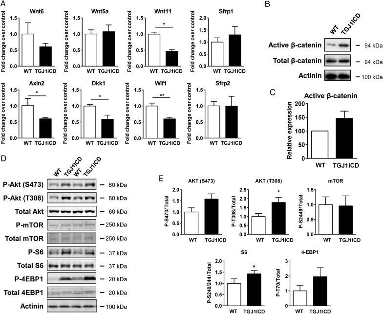
J1ICD-mediated Notch inhibition affects AKT and Wnt signalling pathways. WT and TGJ1ICD neonatal mice were treated daily with tamoxifen until postnatal day P7. (A) Quantitative RT-PCR analysis of Wnt ligands and Wnt inhibitors. Results, normalized to Gapdh, represent fold change relative to WT. (B) The active (dephosphorylated) and the total form of β-catenin were analysed by western blot. An immunoblot anti-α-actinin is shown as a loading control. (C) The active-β-catenin immunoblot was quantified and normalized to total β-catenin. (D) Western blot analysis of AKT and its downstream targets. (E) Bands intensities were quantified by densitometry and the ratios of the phosphorylated to total signals were normalized to the intensity of the WT group. (A, C, and E) Bar graphs are mean ± SEM (n = 5–7 mice per group; *P ≤ 0.05 vs. WT).

