FIGURE 3.
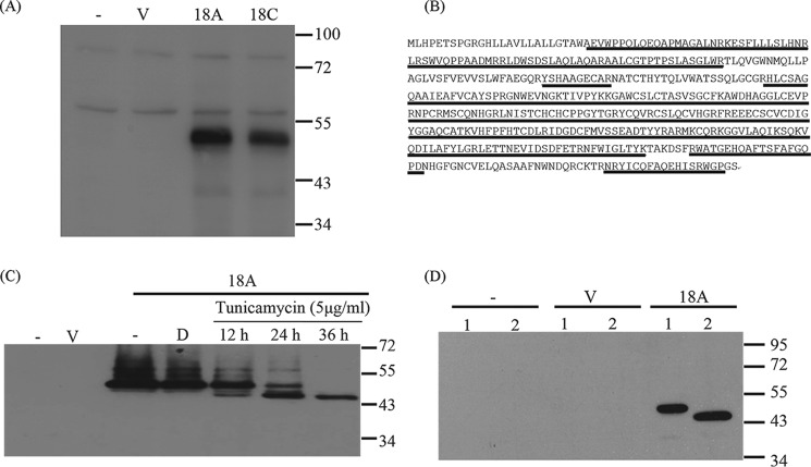
CLEC18 are an N-linked glycoprotein. A, the 293T cells were transfected with pCMV-Tag4A vector (V), pCMV-Tag4A-CLEC18A (18A), or pCMV-Tag4A-CLEC18C (18C), followed by Western blot analysis using anti-CLEC18 mAb (clone 3A9E6). B, mass spectrometry analysis of peptides recognized by anti-CLEC18 mAb (clone 3A9E6). The 50-kDa peptides were eluted for mass spectrometry analysis. The peptides (underlined) matched perfectly to CLEC18. C, CLEC18A-transfected 293T cells were incubated with tunicamycin for various time points, followed by Western blot analysis to determine the presence of N-linked glycosylation using anti-FLAG mAb. D, dimethyl sulfoxide. D, alternatively, 293T cells were transfected with vector (V) or CLEC18A (18A) for 48 h, and then the collected lysate (20 μg) was left untreated (lane 1) or treated with 500 units of peptide:N-glycosidase F (lane 2) for 37 °C for 2 h, followed by Western blot analysis using anti-FLAG mAb.

