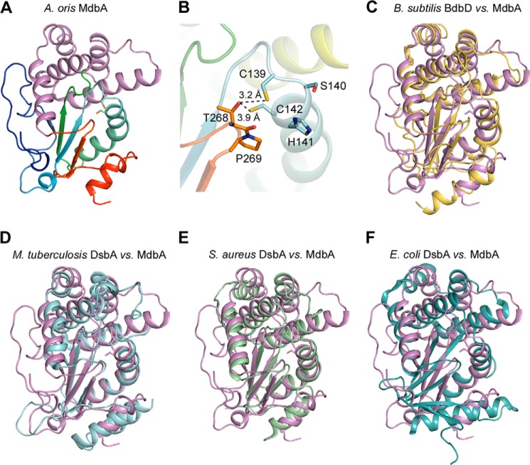
FIGURE 6.
Structural analysis of A. oris MdbA. A, the A. oris MdbA crystal structure (residues 86–296) solved to a 1.55-Å resolution has two domains, a thioredoxin-like domain (rainbow colors) and an α-helical domain (purple). B, the MdbA active site is made of Cys139, Ser140, His141, and Cys142. The Sγ group in the two cysteine residues forms a hydrogen bond with Thr268 of the Cis-Pro element. C–F, the MdbA structure (purple) was aligned with B. subtilis BdbB (C; Protein Data Bank code 3EU3), M. tuberculosis DsbA (D; Protein Data Bank code 4JR6), S. aureus DsbA (E; Protein Data Bank code 3BCI), and E. coli DsbA (F; Protein Data Bank code 1FVK).

