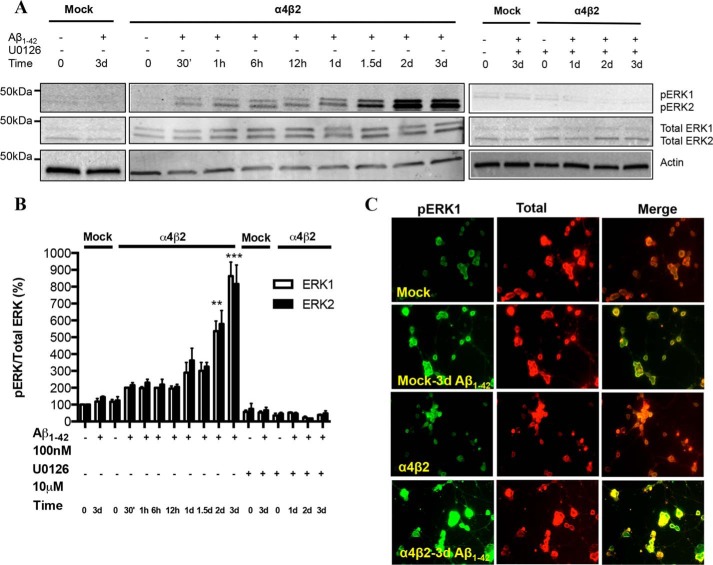
FIGURE 1.
Activation of the ERK MAPK pathway in NG108–15 cells expressing α4β2 nAChRs in response to prolonged exposure to Aβ: time-dependence and sensitivity to a MEK inhibitor. A and B, progressive increases in pERK levels in response to prolonged exposure to 100 nm Aβ1–42 in the absence or presence of U1026, a selective inhibitor of MEK, the upstream regulator of ERK, in cells expressing α4β2-nAChRs or not (Mock: mock-transfected). pERK levels were compared with total ERK as well as actin (loading control). Data are expressed as % (± S.E.) of untreated, mock-transfected controls (n = 3 experiments). C, representative images depicting immunostaining of pERK and total ERK in mock- and α4β2-nAChR-transfected cells treated with Aβ1–42 for 3 days. pERK levels in cells expressing α4β2-nAChRs and treated with Aβ were significantly different in comparison to untreated controls.

