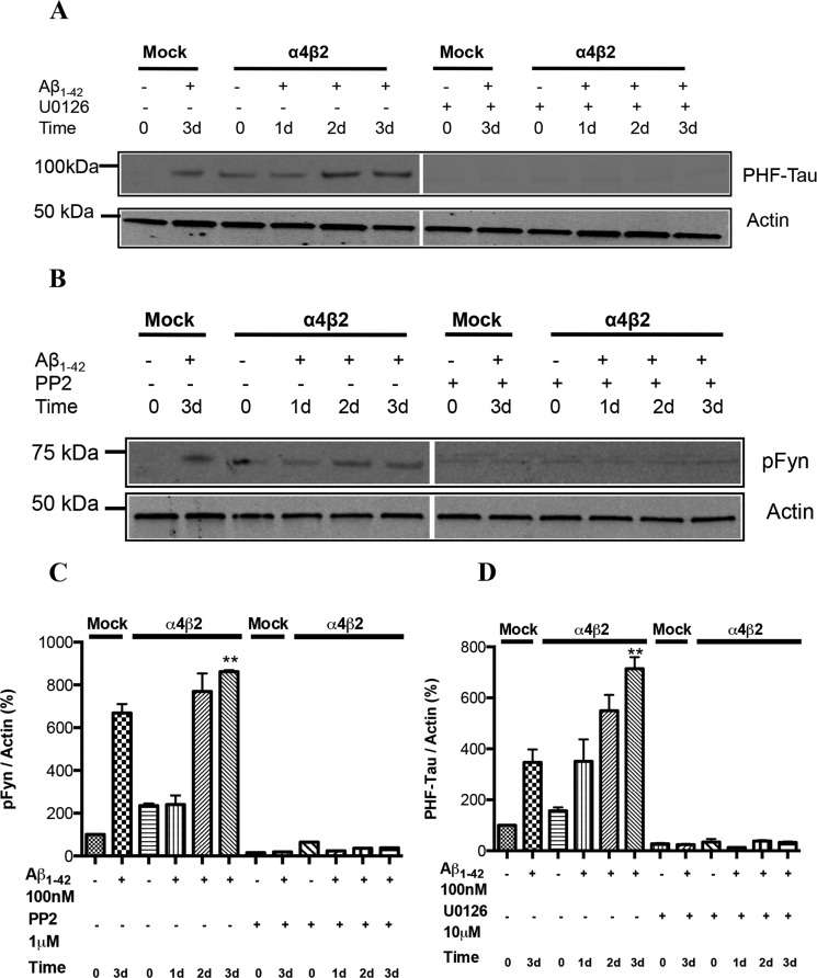
FIGURE 5.
Alterations in the levels of PHF-Tau and pFyn in response to prolonged exposure to Aβ: receptor-dependence. Progressive increases in PHF-Tau (A) and pFyn (B) in differentiated NG108–15 cells expressing α4β2-nAChRs or not (Mock: mock-transfected) response to prolonged exposure to 100 nm Aβ1–42. Note the significant increases following 3-day treatment with Aβ in the absence of target receptor (Mock). Aβ-induced increases in PHF-Tau were compared in the absence or presence of U1026, a selective inhibitor of MEK, while Aβ-induced increases in pFyn were compared in the absence or presence of PP2, a Src kinase family inhibitor. C and D, quantification of averaged immunoblot intensities, as compared with actin (loading control). Data are expressed as % (± S.E.) of untreated, mock-transfected controls (n = 3 experiments). **, p < 0.01 for PHF-tau or pFyn levels in cells expressing α4β2-nAChRs and treated with Aβ for 2 and 3 days when compared with untreated control.

