
FIGURE 12.
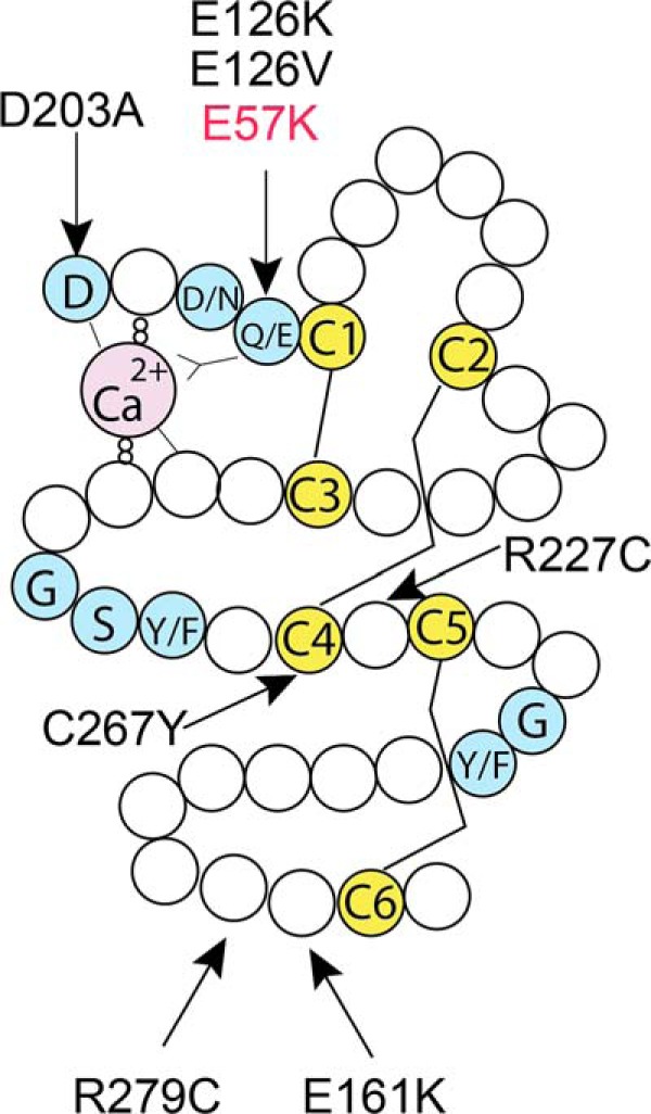
Fibulin-4 missense mutations found in patients. Schematic drawing of a typical cbEGF domain, showing the consensus sequences for calcium binding at the N terminus (blue), six conserved cysteine residues (C1–C6, yellow), and other conserved amino acids (blue). Three pairs of disulfide bridges are formed between C1 and C3, C2 and C4, and C5 and C6. The modified cbEGF domain contains a 28-amino acid insertion between C4 and C5. Arrows show the locations of known fibulin-4 missense mutations within the respective cbEGF domain.