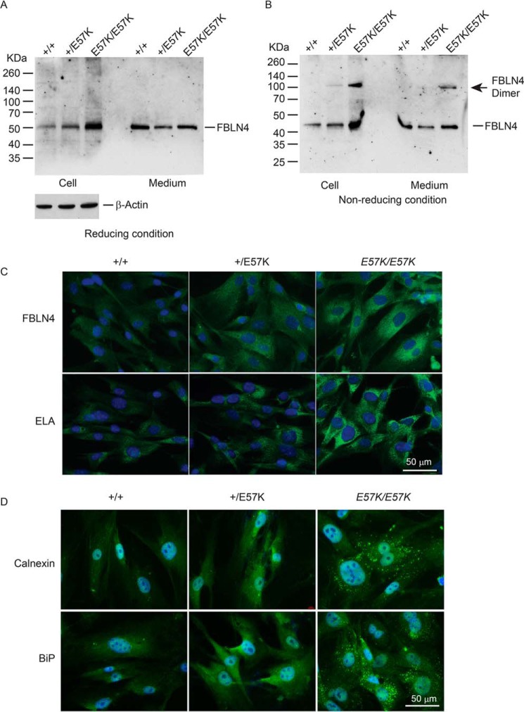
FIGURE 3.
Analyses of dermal fibroblasts from Fbln4+/+, Fbln4+/E57K, and Fbln4E57K/E57K mice. A and B, immunoblot analysis of fibulin-4. Confluent dermal fibroblasts from 4-month-old littermates of three genotypes were grown in serum-free medium for 24 h. Culture medium (100 μl) and cell lysates (20 μg total protein) were separated on 4–12% polyacrylamide gels under reducing (A) and nonreducing (B) conditions. Immunoblot analysis of β-actin in cell lysates (20 μg of total protein/lane) is shown in the bottom of A. C, immunostaining of dermal fibroblasts grown on chamber slides with antibodies against fibulin-4 (FBLN4) and tropoelastin (ELA). Graded increases in intracellular immunoreactivity (green color) of fibulin-4 and tropoelastin were seen in Fbln4+/E57K and Fbln4E57K/E57K fibroblasts, compared with Fbln4+/+ cells. Slides were counterstained with DAPI (blue) to visualize nuclei. D, immunostaining of dermal fibroblasts grown on chamber slides with antibodies against calnexin and BiP. Slides were counterstained with DAPI (blue) to visualize nuclei. Graded increases in immunoreactivity for both ER stress markers were seen in Fbln4+/E57K and Fbln4E57K/E57K fibroblasts, compared with the Fbln4+/+ fibroblasts. Note the punctate staining patterns for both markers in the Fbln4E57K/E57K fibroblasts.

