FIGURE 6.
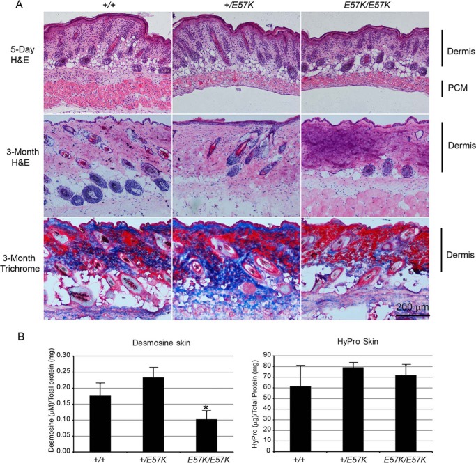
Histological and biochemical analyses of skin from Fbln4+/+, Fbln4+/E57K, and Fbln4E57K/E57K mice. A, top row, cryosections of skin from newborn mice stained with hematoxylin-eosin (H&E), showing thinner dermis and panniculus carnosus muscle (PCM). Middle row, cryosections of skin from 3-month-old mice stained with hematoxylin-eosin, showing a darker purple color in the Fbln4E57K/E57K dermis. Bottom row, cryosections of skin from 3-month-old mice stained with Masson's trichrome reagent showing fewer collagen fibers (blue color) in the Fbln4E57K/E57K dermis. B, desmosine and hydroxylproline analyses of skin from 4-month-old mice (three mice of each genotype). Desmosine content of the Fbln4E57K/E57K skin is significantly reduced compared with the other two genotypes (left panel). The asterisk indicates statistically significant changes (p < 0.05, two-tailed distribution) between knock-in and wild type mice by unpaired Student's t test, assuming equal variances. Hydroxylproline contents of skin from different genotypes are comparable (right panel).

