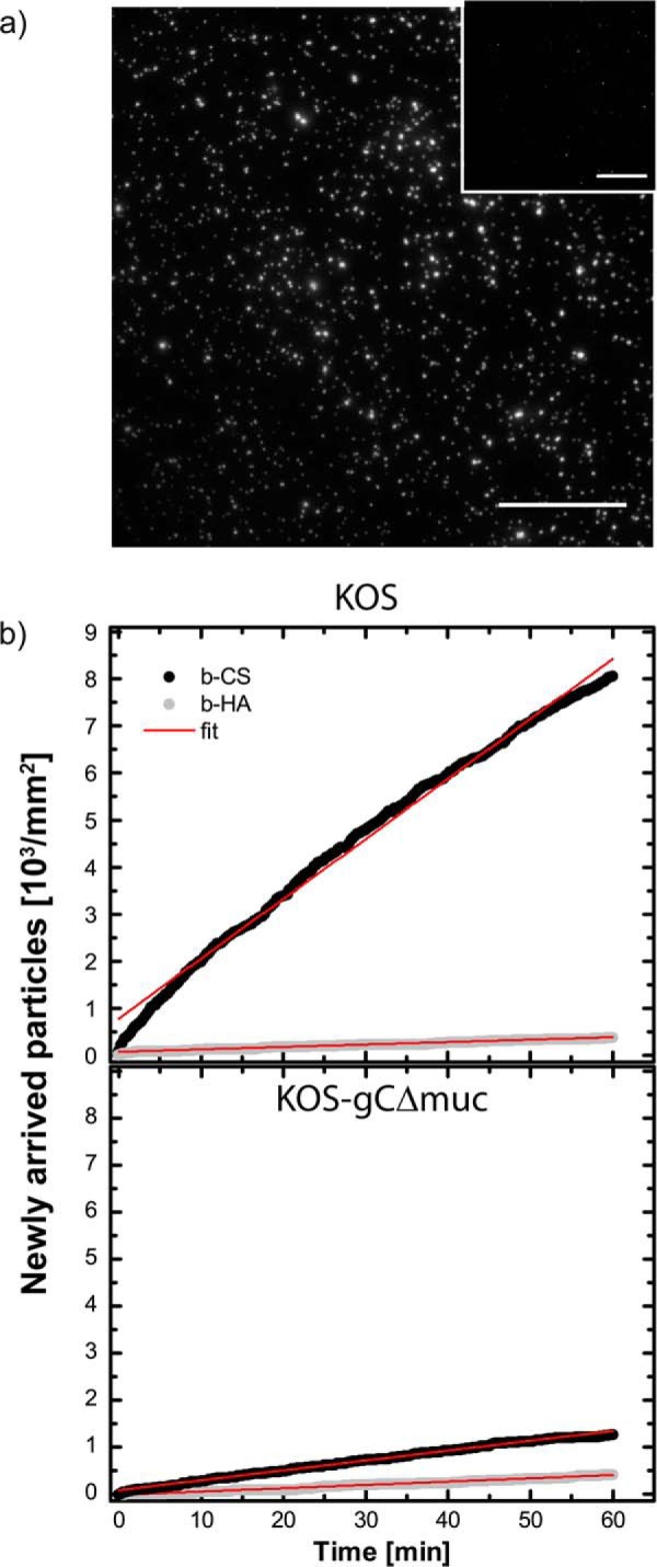
FIGURE 9.

HSV-1 association with surface-bound GAGs investigated by total internal reflection fluorescence microscopy. a, TIRF images showing fluorescently labeled KOS viruses bound to b-CS and b-HA (inset). Scale bar, 50 μm. b, the number of newly arrived virus particles over time was assessed under equilibrium conditions for KOS and KOS-gCΔmuc to b-CS (black dots) and b-HA (gray dots). The data were fitted linearly (red lines).
