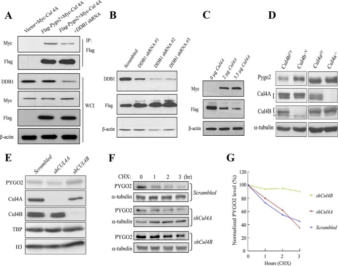
FIGURE 4.
Cul4-DDB1 E3 ligase complex mediates Pygo2/PYGO2 degradation. A, Pygo2 interacts with Cul4A in a DDB1-dependent manner. HEK293T cells were transfected with the indicated constructs with or without DDB1 shRNA, and immunoprecipitation was performed using anti-Myc antibody. B, effect of DDB1 knockdown on Pygo2 level. HEK293T cells that stably express FLAG-Pygo2 were transfected with the indicated shRNAs, and whole cell lysates were used for immunoblotting with the indicated antibodies. C, Cul4A overexpression decreases Pygo2 level. HEK293T cells were transfected with increasing amounts of a Myc-tagged Cul4A-expressing construct, and protein expression was examined 24 h later, as indicated. D, Pygo2 expression in Cul4A−/− or Cul4B−/Y mouse embryonic fibroblasts. Note that the Cul4B gene resides in the X chromosome. The upper bands of the doublets (indicated by brackets) represent neddylated forms. o in the Cul4B panels, nonspecific species. E, effects of Cul4A and Cul4B knockdown on PYGO2 level in BT474 cells. Cells were transfected with the indicated shRNAs, and whole cell lysates were used for immunoblotting with the indicated antibodies. F and G, stabilization of PYGO2 in BT474 cells upon silencing of Cul4B but not Cul4A. The half-lives of PYGO2 were determined in control or BT474 cell lines with stable Cul4A or Cul4B knockdown. Cells were treated with 100 μm CHX for the indicated time period. PYGO2 levels were determined by Western blotting (F), quantified by an Odyssey Infrared imaging system, and normalized to α-tubulin levels (G) at the indicated time points.

