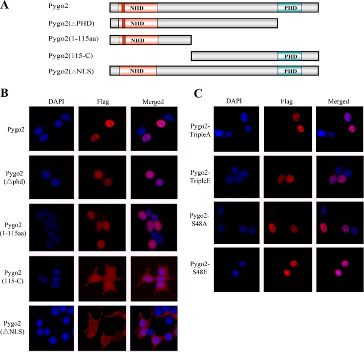
FIGURE 6.
Nuclear localization of Pygo2 deletion mutants. A, schematic diagram of the deletion mutants. B and C, indirect immunofluorescence showing the subcellular localization of FLAG-Pygo2 and its mutant derivatives in HEK293T cells. Cells were transfected with the indicated constructs, and staining was performed 12 h later using anti-FLAG antibody. DAPI stains the nuclei.

