
FIGURE 2.
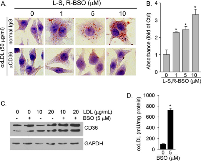
BSO induces macrophage oxLDL uptake by activating CD36 protein expression. A, RAW264.7 cells were treated with BSO for 16 h. After treatment cells were incubated with normal IgG or anti-CD36 antibody (0.3 μg/ml) followed by incubation with oxLDL (50 μg/ml). Cellular oxLDL uptake was determined by Oil Red O staining. B, accumulated Oil Red O dye within cells in each sample of the upper panel in Fig. 2A was extracted individually. The absorbance of extraction solution was determined at 510 nm and normalized by cellular protein content. *, p < 0.05 versus control. C, RAW264.7 cells were treated with LDL or LDL plus BSO as indicated for 16 h. D, RAW264.7 cells were treated with LDL (20 μg/ml) or LDL plus BSO (5 μm) for 16 h. OxLDL concentrations in the cellular lysate were determined by the oxLDL ELISA assay kit. *, p < 0.05 versus LDL alone (n = 3). mU, milliunits.