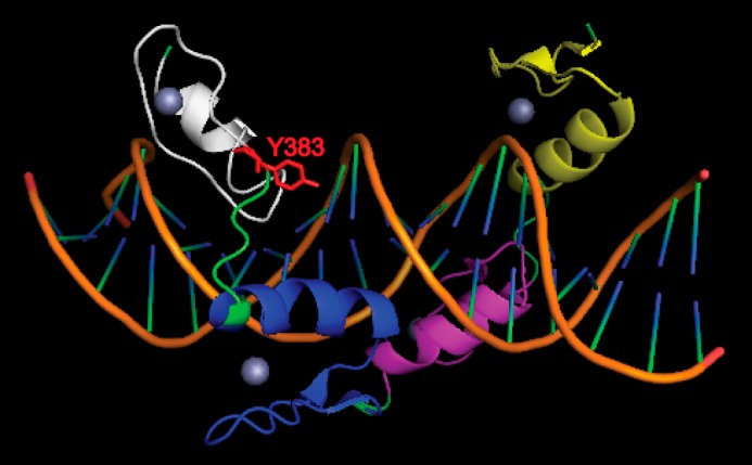
FIGURE 7.

Structure of YY1 DNA binding domain in complex with AAV P5 promoter initiator element. The DNA is shown in orange. The zinc fingers of YY1 are shown as yellow, purple, blue, and white from the N to C termini. Zinc ions are depicted as spheres. The critical tyrosine 383 residue with its side chain is shown in red. Note that structure shown is based on previous report (39).
