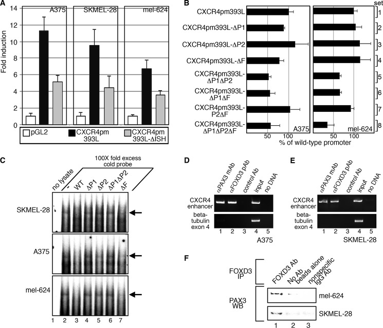
FIGURE 5.
The CXCR4 enhancer is active in melanoma cells through the PAX and FOX sites. A, the CXCR4 intronic enhancer is active in melanoma cells. Empty vector (pGL2) or CXCR4 reporter constructs shown in Fig. 3E were transfected into A375, SKMEL-28, and mel-624 melanoma cells. Luciferase levels are shown as fold-units over controls (levels without PAX3 or FOXD3 expression). B, mutation of PAX and FOX sites attenuates the activity of the CXCR4 enhancer in melanoma cells. A375 and mel-624 cells were transfected with pGL2-CXCR4pm393L constructs with or without mutations in P1, P2, and/or F sites. Luciferase levels are shown as percent of control luciferase levels (pGL2-CXCR4pm393L). C, EMSA analysis using a probe with the CXCR4 enhancer element with melanoma cell lysates produces slow migrating bands. Labeled probe encompassing the CXCR4 enhancer region as shown in Fig. 4B was incubated with SKMEL-28, A375, and mel-624 cell lysates without (lane 2) or with (lanes 3–7) ×100 cold probes with or without PAX or FOX sites mutated as indicated, which may inhibit protein binding to the labeled probe. Lane 1 contains samples with labeled probe without added cell lysate (negative control). Major shift band is indicated with arrows. D and E, PAX3 and FOXD3 are located at the CXCR4 locus in A375 (D) and SKMEL-28 (E) cells. ChIP analysis was performed by precipitating proteins with antibodies specific for PAX3 (lane 1), FOXD3 (lane 2), or with nonspecific normal mouse IgG (lane 3) as a negative control. Additional controls were DNA input (positive control, lane 4) and no template DNA/water blank (negative control, lane 5). Precipitated DNA fragments were utilized as PCR templates with primers specific for the CXCR4 intronic region (top gels) or exon 4 of the β-tubulin gene (bottom gels, negative controls). F, PAX3 and FOXD3 directly interact in melanoma cells. Lysates from mel-624 (top bands) and SKMEL-28 (bottom bands) cells are immunoprecipitated (IP) with a FOXD3-specific antibody (lane 1), no antibody/beads only (lane 2), or with a nonspecific human IgG (lane 3), and probed for PAX3 expression. WB, Western blot.

