FIGURE 2.
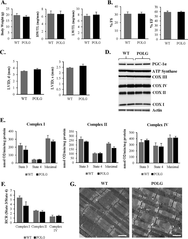
No differences in cardiac and mitochondrial function in WT and POLG mice at 2 months. A, average body weight, heart weight/tibia length (HW/TL), and lung weight/tibia length (LW/TL) ratios were measured in age-matched WT and POLG litter mates (n = 10). Echocardiography measurements at 2 months of age show no significant differences in % fractional shortening (% FS) (B) and % ejection fraction (% EF), left ventricular internal dimension at diastole (LVID; d), and left ventricular internal dimension at systole (LVID; s) (n = 7) (C). D, representative Western blot of mitochondrial biogenesis and OXPHOS proteins in the heart at 2 months. E, mitochondria were isolated from the hearts of 2-month-old mice. Substrates and inhibitors specific to each respiratory complex were added, and oxygen consumption was measured using an oxygen electrode (n = 3). F, respiratory control ratio (RCR) was calculated by dividing state 3/state 4 oxygen consumption. G, transmission electron micrographs show normal mitochondrial ultrastructure in the hearts of WT and POLG mice. Scale bar = 1 μm.

