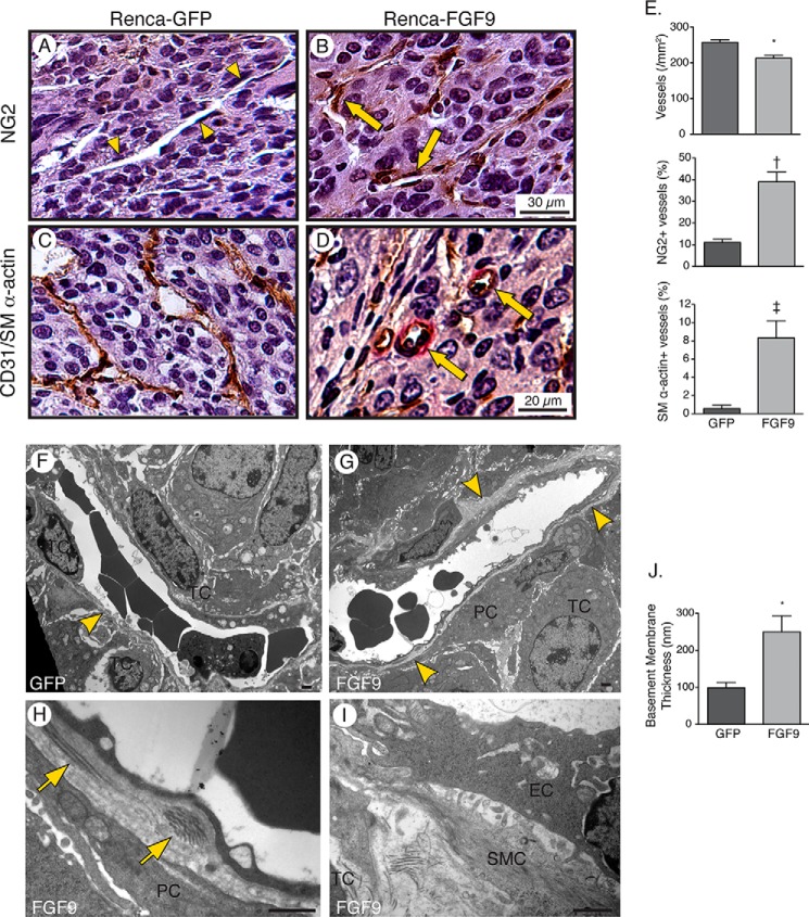
FIGURE 3.
FGF9 stimulates recruitment of vascular mural cells and deposition of a reinforced basement membrane. A–D, photomicrographs of 5-μm Tris-buffered zinc-fixed renal tumor sections harvested 14 days after renal injection of Renca cells expressing GFP and FGF9 and immunostained for NG2-positive pericytes (A and B) or CD31 (brown) and SM α-actin (red) (C and D). Arrowheads depict microvessel devoid of NG2-positive pericytes and arrows depict microvessels invested by pericytes. Some SM α-actin-positive microvessels have an arteriolar morphology (arrows). E, graphs depicting the density of microvessels (*, p = 0.003), percentage of microvessels invested with NG2-postive pericytes (†, p = 0.015), and percentage of microvessels associated with SM α-actin expressing cells (‡, p = 0.002), n = 6. F–I, transmission electron micrographs of microvessels showing an incomplete endothelial basement membrane (BM, arrowhead), with encroaching tumor cells (TC), in a 14-day GFP-expressing tumor (F). A circumferential basement membrane exists in the FGF9-expressing tumor (G, arrowheads) with pericytes (PC) and SMCs separating tumor cells from the vessel (G–I). Arrows depict fibrillar collagen deposits in the basement membrane. EC, endothelial cell. Bar, 1 μm. J, graph depicting the mean thickness of the basement membrane of tumor microvessels, measured from 25 microvessels in GFP-expressing tumors and 20 vessels in FGF9-expressing tumors (*, p = 0.017).

