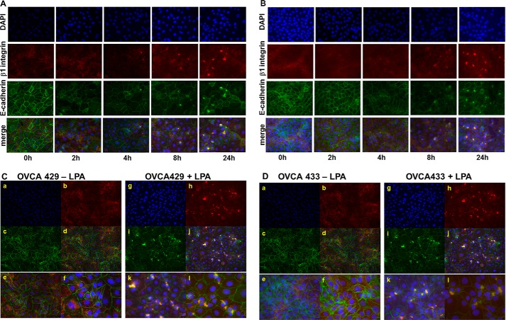
FIGURE 4.
LPA induces β1 integrin aggregation and β1 integrin/E-cadherin co-localization. A and B, OVCA 429 (A) or OVCA433 (B) cells were grown on coverslips coated with type I collagen (10 μg/ml) prior to treatment with LPA (30 μm) for the time periods shown. Cells were stained to evaluate localization of β1 integrin (anti-β1 integrin, 1:50 and Alexa Fluor 594 goat anti-mouse IgG, 1:400, red) or E-cadherin (anti-E-cadherin, 1:100 and Alexa Fluor 488 goat anti-rabbit IgG, 1:400, green). Nuclei are stained with DAPI (blue). Merged images (×40 magnification) show β1 integrin clustering beginning at 4 h. C and D, co-localization of E-cadherin with clustered β1 integrin. OVCA 429 (C) or OVCA433 (D) cells were grown in the absence (left panels) or presence (right panels) of LPA (30 μm) for 24 h prior to analysis by dual label immunofluorescence microscopy. Panels a and g, DAPI; panels b and h, β1 integrin; panels c and i, E-cadherin; panels d–f and j–l, merged images. Panels a–d and g–j, ×20 magnification; panels e and k, ×40 magnification; panels f and l, ×60 magnification. Merged images demonstrate LPA-dependent co-localization of E-cadherin with clustered β1 integrins. Areas of co-localization of E-cadherin with β1 integrin are visible in cells treated with LPA treatment (right panels, C and D) relative to untreated controls (left panels, C and D).

