
FIGURE 5.
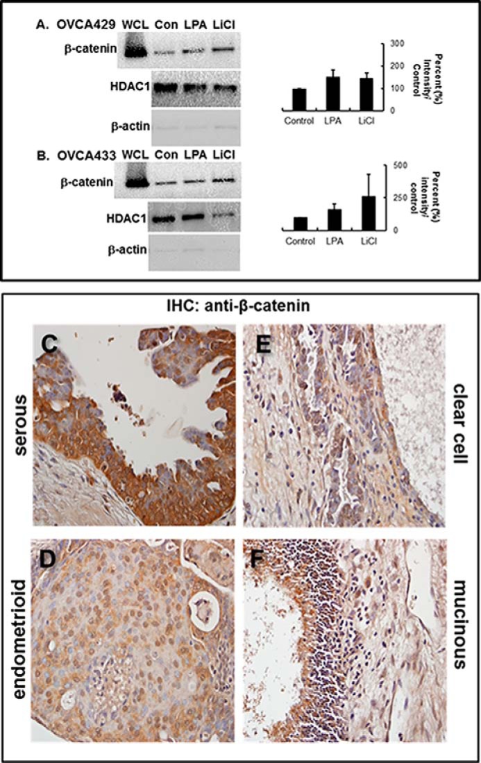
LPA induces LPA receptor-dependent nuclear translocation of β-catenin. A and B, cells were treated as labeled (Con, untreated control; LPA, 40 μm; LiCl, positive control, 40 μm) and then subjected to subcellular fractionation as described under “Experimental Procedures.” Fractions were electrophoresed on 9% SDS-polyacrylamide gels and immunoblotted for β-catenin expression (anti-β-catenin, 1:1000). From analysis of triplicate blots, β-catenin staining in the nuclear fraction of both OVCA429 (A) and OVCA433 (B) cell lines was increased ∼50% in LPA-treated cells compared with control (graphs). Control blots were probed with HDAC1 (middle panels) or β-actin (lower panels) to detect the presence of nuclear or cytoplasmic compartments, respectively. WCL designates unfractionated whole cell lysate. Molecular mass of β-catenin, 92 kDa. C–F, representative samples of each of the four major EOC histotypes were stained by immunohistochemistry (IHC) with anti-β-catenin (BD Transduction Laboratories, 1:50) and a biotinylated secondary antibody (1:200; Vectastain ABC, Vector Laboratories). Slides were finally subjected to 3,3′-diaminobenzidine peroxidase (Vector Laboratories) exposure and hematoxylin staining. β-Catenin staining is present at cell-cell junctions, in the cytoplasm, and in the nucleus. Nuclear β-catenin is found in serous (C), endometrioid (D), clear cell (E), and mucinous (F) tumors.