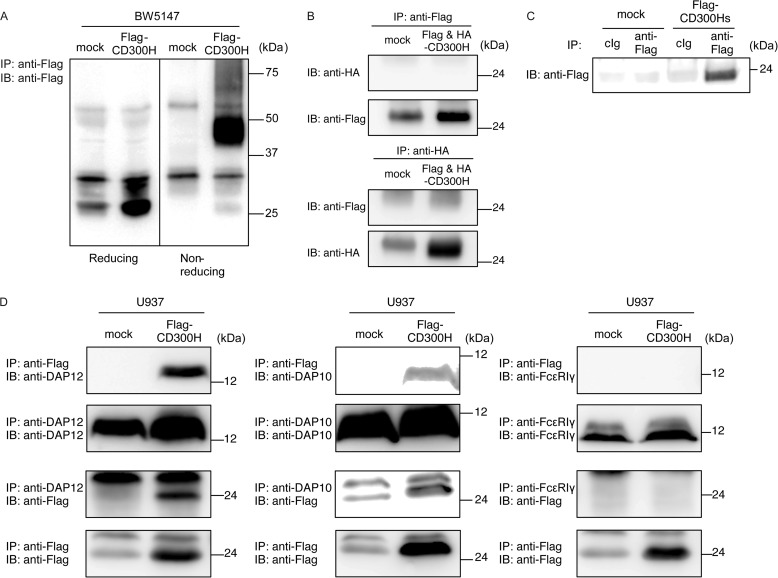
FIGURE 5.
Biochemical analyses of CD300H. A, BW5147 cells and transfectants expressing FLAG-tagged CD300H (5 × 10⁶ cells/experiment) were lysed in 1% Nonidet P-40 buffer, immunoprecipitated (IP) with anti-FLAG, and immunoblotted (IB) with anti-FLAG. B, BW5147 transfectants simultaneously expressing FLAG-CD300H and HA-CD300H were lysed in digitonin buffer, immunoprecipitated with anti-HA or anti-FLAG, and immunoblotted with anti-HA or anti-FLAG in reducing conditions. C, culture supernatant from 293T cells transiently expressing FLAG-tagged CD300Hs or mock were immunoprecipitated with anti-FLAG and immunoblotted with anti-FLAG. D, U937 cells and transfectants stably expressing FLAG-tagged CD300H were lysed in digitonin buffer, immunoprecipitated with anti-FLAG, anti-DAP12, anti-DAP10, or anti-FcϵRIγ, and immunoblotted with anti-FLAG, anti-DAP12, anti-DAP10, or anti-FcϵRIγ. Data are representative of two independent experiments.

