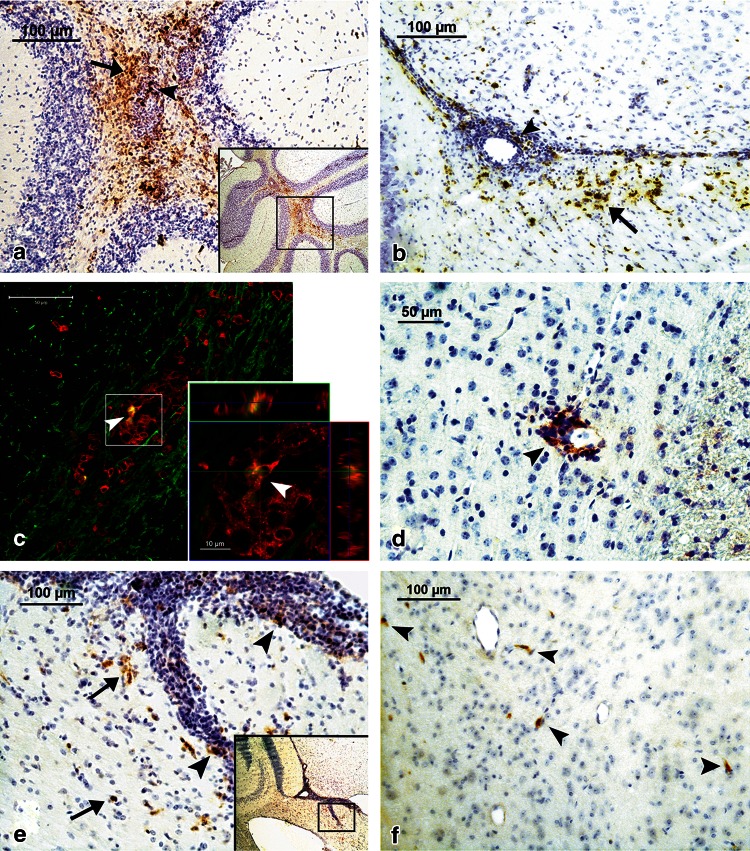
Fig. 3.
Immunohistochemical analysis of expression of CCL19 (panels a, b, d), CCL21 (panels e, f) and colocalization of CCL19, CD3 and MBP antigens (panel c) in the brain during ChREAE. a CCL19 is expressed abundantly during the first attack of ChREAE in many areas of the brain including the cerebellum (inset). CCL19-positive cells, most of them displaying a morphology of mononuclear leukocytes are present within perivascular inflammatory cuffs (arrowhead). A strong staining is also present in surrounding infiltrated parenchyma (arrow). b During the first attack of disease the expression of CCL19 was detected also in the infiltrated periventricular white matter in the vicinity of inflammatory lesions (arrows). In these inflammatory lesions expression of CCL19 was also present (arrowhead). c CCL19 (yellow) and CD3 (red) are expressed in the same inflammatory lesion in the periventricular white matter (myelin basic protein, MBP—green). A high magnification of this lesion shows (arrowheads) that expression of CCL19 (yellow) is colocalized with some cells expressing CD3 (red). d During chronic phase of the disease expression CCL19 in the brain was also detected using immunohistochemistry but in much lesser degree near only a small number of inflammatory lesions. e CCL21 expression was observed during the first attack in many areas of the brain also near the lateral ventricles (inset). Higher magnification shows that positive staining was present both within (arrowheads) and near (arrows) inflammatory cuffs, in the infiltrated brain parenchyma. f During the chronic phase of the disease CCL21 staining still could be detected but only few positive cells were observed in brain parenchyma in the vicinity of blood vessels (arrowheads). The immunostaining procedure was performed according to the protocols described in “Materials and methods”

