Figure 5.
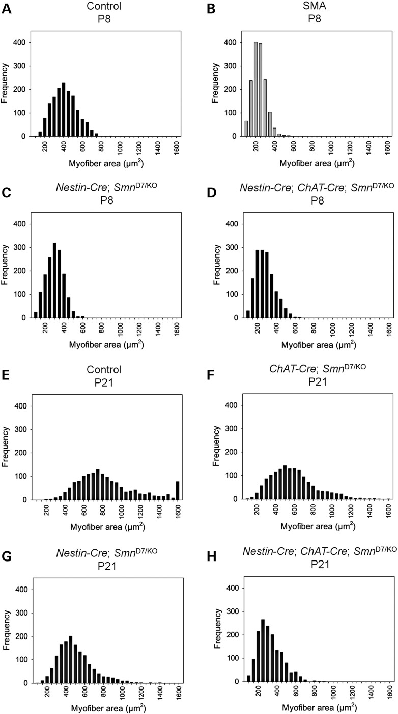
Muscle fiber size upon deletion with Nestin-Cre and ChAT-Cre. The gastrocnemius muscle from (A) control, (B) SMA, (C) Nestin-Cre Smn-deletion mice and (D) Nestin-Cre + ChAT-Cre Smn-deletion mice were measured at P8. Myofiber size in control mice at P8 (mean 387.8 ± 3.3 µm2; median 379.5 µm2) is significantly different from that of Nestin-Cre Smn-deletion mice (mean 277.5 ± 2.3 µm2; median 277.0 µm2), Nestin-Cre + ChAT-Cre Smn-deletion mice (mean 257.4 ± 2.5 µm2; median 247.0 µm2, P < 0.001). The Nestin-Cre + ChAT-Cre Smn-deletion mice however have myofiber areas similar to that of SMNΔ7 SMA mice at P8 (Mean 210.6 ± 1.9 µm2; median 205.1 µm2) (51). (E–H) The difference in myofiber area is more dramatic at P21 where (E) control mice (mean 835.9 ± 8.8 µm2; median 757.0 µm2) and (F) ChAT-Cre deletion mice (mean 559.6 ± 6.1 µm2, median 530.5 µm2) display larger fibers as compared with (G) Nestin-Cre deletion mice (mean 477.6 ± 4.8 µm2, median 447.0 µm2) and (H) Nestin-Cre + ChAT-Cre Smn-deletion mice (mean 302.7 ± 3.2 µm2; median 281.0 µm2, P < 0.001). There is no comparison to SMNΔ7 SMA mice at this time point because the SMNΔ7 SMA mice only live for 14 days. For each group, a total of 1500 fibers were measured from 3 mice (approx. 500 fibers per mouse).

