
Figure 1.
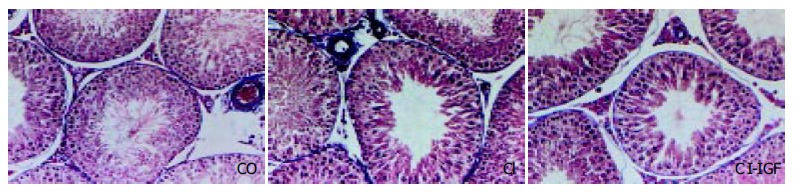
Microscopy of testes (× 150 magnification, Masson’s stain). Testicular histological sections of normal rat (CO) demonstrated active spermatogenesis in normal-size seminiferous tubuli with thin basement membranes and minimal peritubular fibrosis. Leydig cells were scarce, being widely separated by seminiferous tubuli. No evidence of peritubular fibrosis and other alterations were found in testes from cirrhotic animals.