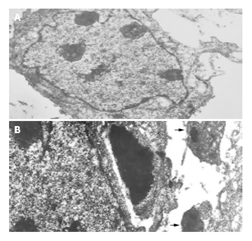
Figure 2.

Transmission electron microscopic photographs of the SGC7901 cells cultured for 96 h in the absence (A) or the presence of 20 μmol/L mifepristone (B) in vitro (TEM, × 2000). Arrows indicate apoptotic bodies which were formed in the cells of mifepristone-treated group.
