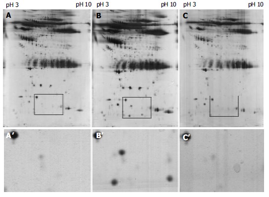
Figure 2.

Representative of two-dimensional electrophoresis patterns of sera protein from the same esophageal squamous cell carcinoma patient before (A) and after (B) operation and the normal subject (C) with different acidity from pH3 to pH10. A’, B’ and C’ are the magnification of A, B and C, respectively, for the sera protein with marked difference.
