
Figure 1.
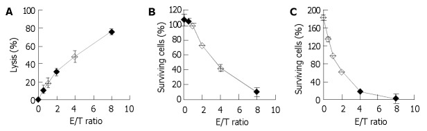
Dose-response relationships of 51Cr release assay and the two crystal violet staining assays. PBMCs were detected after 15-d culture with irradiated HFWT cells. A: 51Cr release assay, in which the effector lymphocytes and the fresh target cells pre-labeled with 51Cr were incubated for 4 h. B and C: crystal violet staining, in which effector lymphocytes and fresh target HFWT cells were incubated for 4 and 24 h, respectively. Each point and bar represent mean and SD (3-6 replicates), respectively.