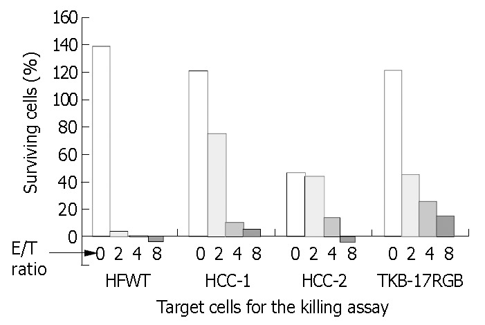
Figure 3.

Killing activity of the NK-enriched population on MHC class I-positive and -negative cell lines. PBMCs of HCC patient were expanded on irradiated T cells and submitted to the 24-h crystal violet staining. Target cells were MHC class I-negative HFWT and HCC-2 cells, MHC class I-positive HCC- 1 and TKB-17 RGB cells. HCC-1 and HCC-2 cells were derived from the same human hepatocellular carcinoma tissue.
