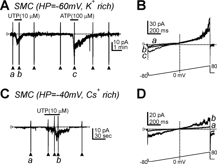
Fig. 3.
Effects of UTP on membrane currents in smooth muscle cells (SMCs). A: UTP (10 μM) and ATP (100 μM) induced inward currents in SMCs dialyzed with a K+-rich solution at a HP of −60 mV. B: expanded timescales from control conditios (a) and in the presence of UTP (b) in A. C: UTP activated inward currents in SMCs dialyzed with Cs+-rich pipette solution (HP = −40 mV). D: expanded timescales from before (a) and after UTP (b) in C. Horizontal dotted lines denote 0 pA. Arrowheads denote the applications of ramp potentials (protocol shown in bottom traces in A and C).

