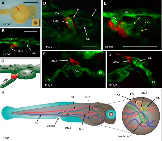
Fig. 1.
Localization of the principal site of renin gene (ren) expression. A: dorsal view of ren mRNA within the location of the anterior mesenteric artery (AMA). The lumen is visible in the inset. Scale bar = 500 μm. hpf, hours postfertilization. B: ren-red fluorescent protein (RFP) in Tg(ren:RFP, kdrl:GFP, casper; where GFP is green fluorescent protein) becomes first visible at ∼48 hpf at the origin of the AMA, where it branches off the ventral dorsal artery (DA) immediately caudal to the glomerular primordium (GL). The exact timing of ren-RFP expression varies between individuals. Scale bar = 25 μm. C: schematic of the image in B showing renin expression (red), the AMA, DA, and GL endothelium (green), and blood flow (arrows). D: Tg(ren:RFP, casper) and FITC-dextrans showing mural ren-RFP and a patent vasculature. Afferent arterioles entering the GL (a) and an efferent arteriole draining into the AMA at 72 hpf (b) are shown. Scale bar = 50 μm. E: multiphoton projection showing details of mural cell ren-RFP and kdrl-GFP at the AMA in Tg(ren:RFP, kdrl:GFP, casper) at 96 hpf. Yellow arrows show two glomerular arterioles draining into the AMA. Scale bar = 50 μm. F and G: dorsal (F) and sagittal (G) views of Tg(ren:RFP, wt1b:GFP, casper) at 96 hpf showing juxtaglomerular ren-RFP at the AMA. At 96 hpf, faint ren-RFP expression was visible on the ventral DA immediately caudal to the AMA (red arrow). Developing proximal tubules (PTs) also express Wilms' tumor 1b (wt1b)-GFP. Scale bars = 50 μm. H: schematic showing sites of renin expression at 5 days postfertization (dpf). Mural renin cells are shown in yellow, arterial vessels are red, venous vessels are blue, and the GL, nephron, and cloaca are in green. SIA, supraintestinal artery.

