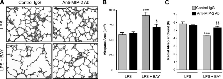
Fig. 5.
MIP-2 neutralization prevents the severe disruption of alveolarization induced by inhibiting the NF-κB pathway in the LPS-exposed lung. A: representative images of H&E-stained lung sections from neonatal mice pretreated with either control IgG or anti-MIP-2 Ab, obtained 24 h after administration of LPS or LPS + BAY. Scale bar = 100 μm. B: morphometric analysis to determine the distal airspace area, with ***P < 0.01 vs. LPS + IgG and §P < 0.05 vs. LPS + BAY + IgG. Data are expressed as means ± SE, with n = 5–7 for each group. C: morphometric analysis to determine the radial alveolar count, with ***P < 0.01 vs. LPS + IgG and §§P < 0.01 vs. LPS + BAY + IgG. Data are expressed as means ± SE, with n = 6–8 for each group.

