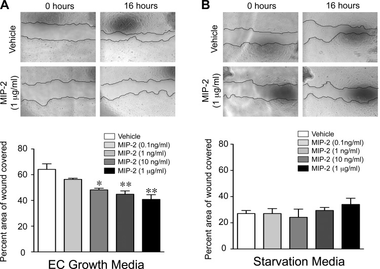
Fig. 7.
MIP-2 directly impairs pulmonary endothelial cell (PEC) migration. Representative phase contrast images of wound made in confluent monolayers of neonatal primary PEC at time 0 h and 16 h, after incubation with vehicle or increasing concentrations of recombinant MIP-2 in endothelial growth media (A) or starvation media (B), are shown. The percent area of the wound covered at 16 h was quantified, with *P < 0.05 and **P < 0.01 vs. vehicle. Data are expressed as means ± SE, with n = 3 for each group.

