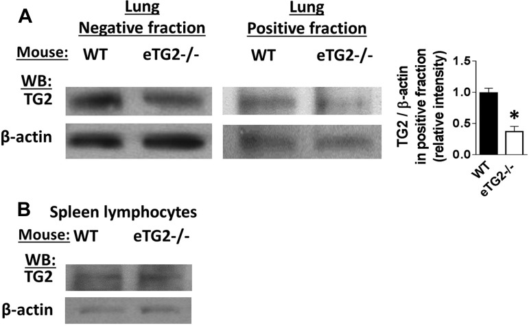
Fig. 2.
TG2 expression in lung endothelial and spleen cells. A: lung endothelial cells were immunomagnetically selected using anti-PECAM-1 and anti-endoglin antibodies. Fractions were analyzed by flow cytometry, and the positive fraction was >80% endothelial cells (data not shown). The negative fraction is the flowthrough from the immunomagnetic column. Left: representative Western blots from a WT and eTG2−/− mouse for TG2 and β-actin for the endothelial cell-positive fraction and the column flowthrough (negative fraction). The light band in the positive fraction from the eTG2−/− mice is due to the 80% purity of the endothelial cells. Right: relative intensity of TG2-to-β-actin ratio in the positive fraction from the immunomagnetic cells, which contained >80% endothelial cells. Values are means ± SE. *P < 0.05. B: spleen cells are >90% lymphocytes and were examined for TG2 expression and β-actin by Western blotting.

