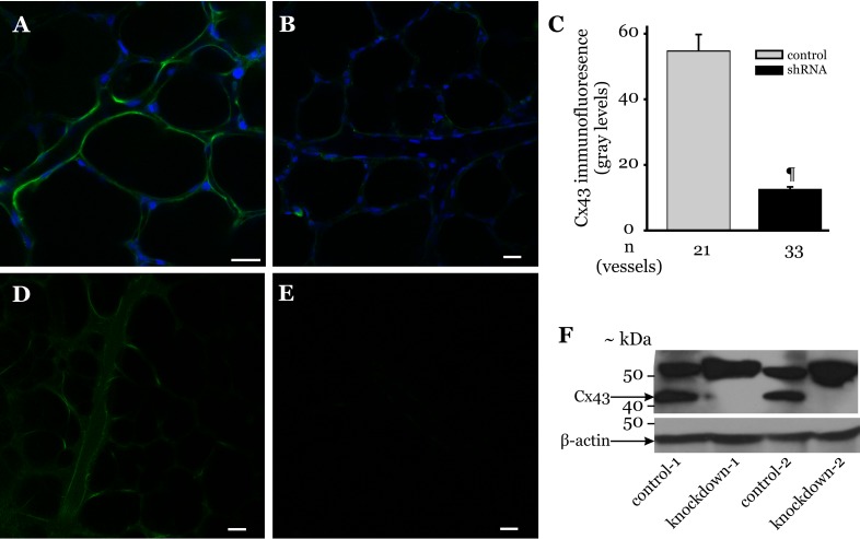
Fig. 5.
Knockdown of Cx43 in intact lung microvessels. A and B: rats were injected with either Cx43 shRNA or vehicle, as outlined in materials and methods. Microvessel Cx43 expression in lungs isolated from these animals was determined by in situ immunofluorescence. Immunofluorescence images show Cx43 expression (green) in vehicle- (A) and Cx43 shRNA-treated lung microvessels (B). Blue staining indicates nuclei. Bar, 20 μm. C: bar graph shows Cx43 expression quantified along margins of microvessels within an image field (>3 images/treatment group were analyzed). n = No. of vessels analyzed. ¶P < 0.001 compared with control treatment (3 lungs/treatment group). D and E: rats injected with P-lenti green fluorescent protein (GFP) lentiviral vector or vehicle, as outlined in materials and methods. Image shows GFP fluorescence in microvessels of lungs isolated from vector- (D) and vehicle-treated rats (E) repeated in 3 animals/treatment group. F: rats were injected with Cx43 shRNA lentiviral vector (1 × 10⁹ TU/ml; 20 μl) and ViraDuctin (2 μl in 100 μl PBS) via the tail vein and allowed to recover for 72 h. Control rats were injected with ViraDuctin (2 μl in 100 μl PBS). After 72 h, the lungs were isolated and the peripheral regions of the lungs dissected and homogenized. The homogenized lung tissue was then prepared for immunoblot analysis of the indicated proteins, as outlined in materials and methods. The Cx43 antibody used in this study also detected a nonspecific band at ∼52 kDa; 2 rats/treatment protocol (indicated by the no. 1 or 2 in the figure). Each lane represents data from 1 rat.

