Fig. 3.
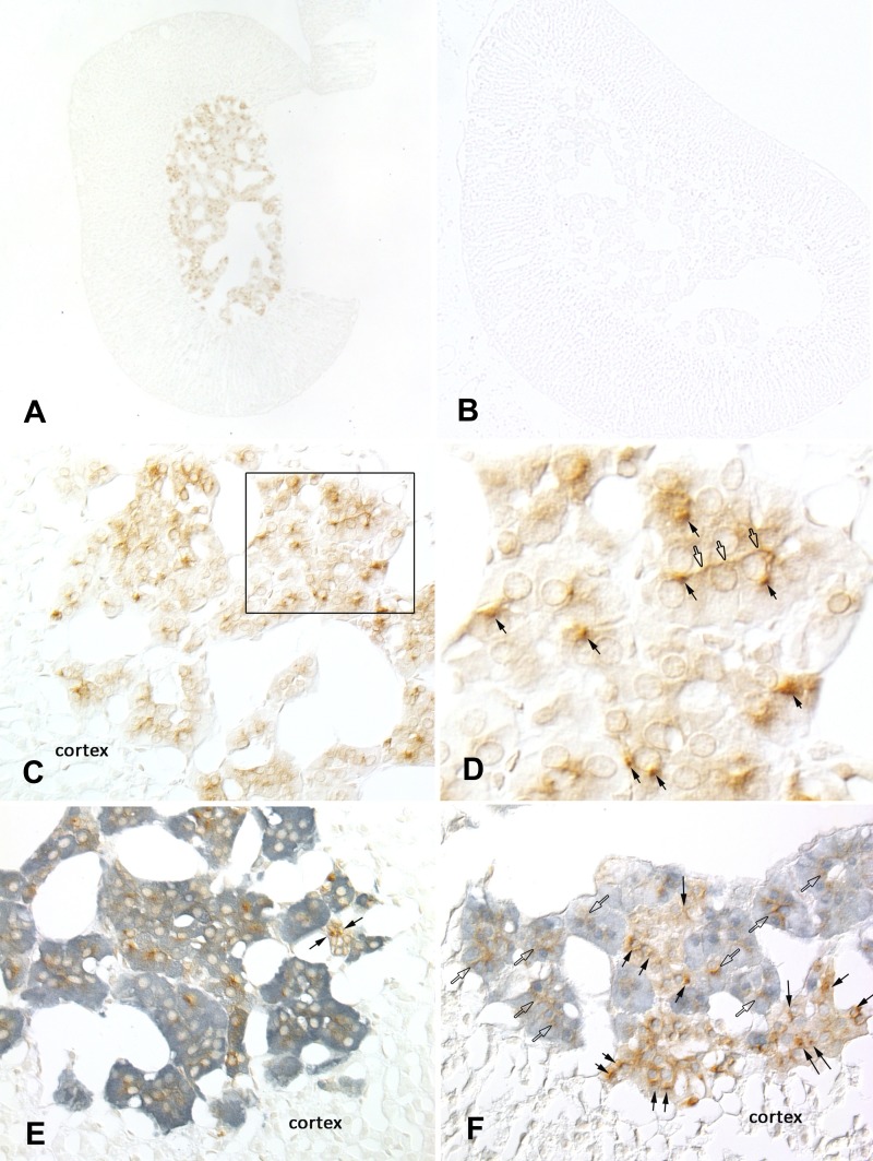
Pendrin is expressed in chromaffin cells of the mouse adrenal medulla. Pendrin immunolabeling was observed in the adrenal medulla of wild-type (A), but not of pendrin-null mice (B). C: pendrin immunolabel in adrenal medulla of wild-type mice. D: inset from C at higher magnification. Open arrows mark areas of peripheral immunoreactivity consistent with plasma membrane labeling; closed arrows show cells with intense foci of immunolabeling within the cell cytoplasm. E: pendrin (brown) and tyrosine hydroxlase (TH; blue) double labeling. Virtually all pendrin-positive cells are also positive for TH. Occasional cells are positive for pendrin, but TH negative (arrows). F: pendrin (brown) and α-phenylethanolamine N-methyltransferase (α-PNMT; blue) double labeling. Pendrin immunoreactivity is present in both α-PNMT-positive (open arrows) and α-PNMT-negative cells (closed arrows). None of these images showed pendrin immunolabel in the adrenal cortex.

