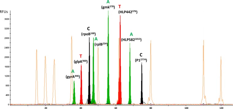
FIG 3.
Example of electropherogram obtained for a M. pneumoniae isolate (SNP type 2) using the eight-plex SNaPshot minisequencing assay (Applied Biosystems). The x axis represents the size (in nucleotides) of the minisequencing products relative to the GeneScan-120 LIZ internal size standard represented with the orange peaks (Applied Biosystems), whereas the y axis represents the relative fluorescence units (RFUs). Each plot was obtained using GeneMapper software (v.4.1; Applied Biosystems). For SNPs glpK360, rpoB168, and HLP442376, the genes were localized on the negative (−) DNA strand, but the SBE primers were designed on the positive (+) DNA strand. For SNP P12774, the gene is localized on the positive (+) DNA strand, but the SBE primer was designed on the negative (−) DNA strand.

