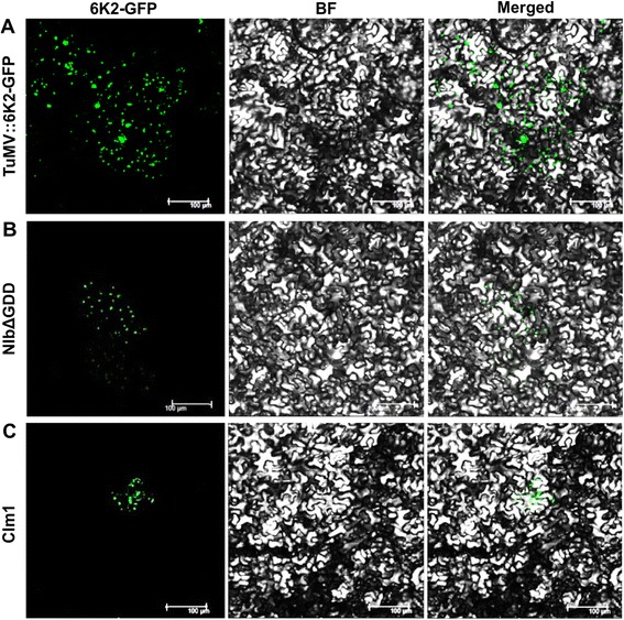
Fig. 2.

Analysis of viral cell-to-cell movement by confocal microscopy. Leaves of Nicotiana benthamina agro-infiltrated with the wild type parental virus TuMV::6 K2-GFP (positive control), the replication-defective mutant NIbΔGDD (negative control) and CI mutants were observed under a confocal microscope 4 days post infiltration. a Multicellular infection of TuMV::6 K2-GFP in the inoculated leaf. b Single cell expression of the replication-defective mutant NIbΔGDD (resulting from the 35S promoter). c Single cell infection of the TuMV::6 K2-GFP CIm1 mutant. Scale bar, 100 μm
