
Fig. 6.
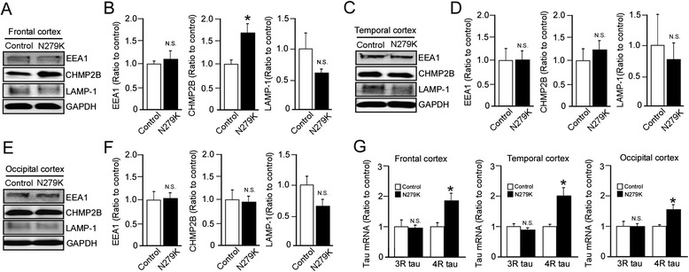
Subcellular vesicle components and tau mRNA levels in the cortices of PPND/FTDP-17 patients with N279K tau mutation. a-f Western blot analysis of EEA1, CHMP2B and LAMP-1 levels in the frontal a, b, temporal c, d and occipital e, f cortex from non-demented controls (n = 4) and PPND/FTDP-17 patients (n = 4). g Quantitative RT-PCR for levels of 3R-tau and 4R-tau mRNA in the frontal, temporal and occipital cortex from control non-demented controls (n = 4) and PPND/FTDP-17 patients (n = 4). Relative gene expression represents fold changes relative to control patient levels, normalized to β-actin. Data are mean ± SEM. *p < 0.05. N.S, Not Significant