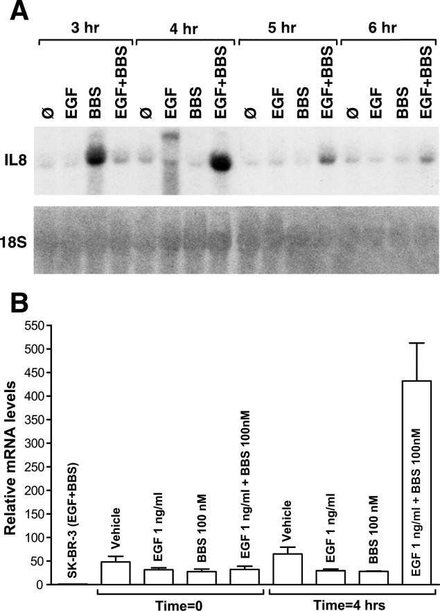
Figure 3.
Time course of IL-8 mRNA induction by EGF and /or BBS. A) Autoradiography of Northern blots demonstrates steady state levels of IL-8 mRNA after treatment with EGF (1 ng/ml), BBS (100nM), or both EGF and BBS. Total RNA (15 μg/lane) was isolated from MDA-MB-231 cells at the indicated time points after treatment. RNA was resolved on a 1% agarose-formaldehyde gel, blotted onto a Hybond-N+ membrane, and hybridized with a [α32P]dATP-labeled cDNA probe. Equal loading and transfer was verified by rehybridizing the blot with a radiolabeled probe for 18S RNAse. B) Quantitative real-time PCR analysis of IL-8 mRNA levels showing synergistic increase at 4 h. As a negative control, mRNA from SK-BR-3 cells was used.

