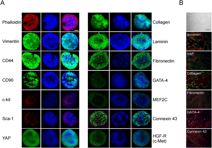
Fig 2. Immunophenotype of hCPC-derived spheroids.
(A) Confocal microscopy images of spheroids for mesenchymal/stromal, stemness, extracellular matrix, and cardiomyogenic markers, YAP and HGF receptor expression using specific antibodies followed by secondary FITC-labelled antibodies or TRITC-phalloidin. One representative out of the three experiments performed with similar results is shown. Scale bar, 50 μm. (B) For comparison, images of hCPCs kept as monolayers at phase contrast microscope (top; scale bar 100 μm) or in immunofluorescence after staining for some of the same markers are shown (20 x magnification).

